

EVALUATION OF BASAL SPHENOID ANGLE AMONG ADULTS OF AFRICAN DESCENT USING TOMOGRAPHIC IMAGING
Article Sidebar
Main Article Content
Abstract
Background: The Basal Angle (BA) of the sphenoid represents an integral anthropological component of the radiological examination of the cranial floor which allows for the diagnosis of platybasia. As a developmental anomaly of the skull base, occipital bone and upper cervical vertebrae, platybasia is associated with changes in the depth of the posterior cranial fossa and the shape of the spheno-occipital region.
Objective: To determine the average basal sphenoid angle and its relationship with the dimensions of the sphenoid sinus among adults of African origin using computerized tomography (CT).
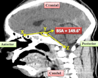
Method: Computerized tomography (CT) images of three hundred and twenty-three adult sphenoid sinuses of individuals with age ranging from 18 to 80 years, over a 5- year period were retrospectively studied at the Radiology Department of the University Teaching Hospital following institutional ethical approval. On a sagittal CT slide, the sphenoid BA was measured as the angle subtended between two lines on the cranial floor. Platybsia was defined as; BA greater than 143°. The anteroposterior, craniocaudal, and transverse diameters of the sphenoid sinus were measured, the extent of pneumatisation was noted, while sinus volume was calculated.
Results: The BA of the sphenoid ranged from 88° to 157.9°with a mean BA of 124.70° ± 11.4. There was a statistically significant relationship between the BA and the craniocaudal diameter of the sphenoid sinus.
Conclusions: Computerized tomography assessment of the sphenoid basal angle among Africans is lower than currently known. These values are also lower compared to results obtained using X-rays and among Caucasians. This emphasises the need for more studies on CT assessment of the skull base for the determination of an applicable African average for the sphenoid BA. This is relevant in the radiological identification of skull base malformations.
Downloads
Article Details
Section

This work is licensed under a Creative Commons Attribution-NonCommercial 4.0 International License.
All articles in JRRS are published under the Creative Commons Attribution 4.0 International License (CC BY 4.0). This permits unrestricted use, distribution, and reproduction in any medium, provided the original work is properly cited.
How to Cite
References
1. Chamberlain WE. Basilar impression [platybasia]: A bizarre developmental anomaly of the occipital bone and upper cervical spine with striking and misleading neurologic manifestations. Yale J Biol Med 1939; 11:487-489.
2. Ray BS. Platybasia with involvement of the central nervous system. Ann Surg 1942; 116:231.
3. Poppel MH, Jacobson HG, Duff BK, et al. Basilar Impression and Platybasia in Paget's disease. Radiology 1953; 61:639-644
4. Adam AM. Skull Radiograph Measurements of Normals and Patients with Basilar Impression; use of Landzert's Angle. Surg Radiol Anat 1987;9: 225-229
5. Scoville WB, Sherman IJ. Platybasia: Report of ten cases with comments on familial tendency, a special diagnostic sign, and the end results of operation. Ann Surg 1951; 133:496.
6. Nachmani A, Aizenbud D, Berger G, et al. The prevalence of platybasia in patients with velopharyngeal incompetence. Cleft Palate Craniofac J 2013; 50:528-534.
7. Ramsey RG. Neuroradiology. 3rd Ed. Philadelphia: WB Saunders 1994; 8: 570–571
8. Koenigsberg RA, Vakil N, Hong TA, et al. Evaluation of Platybasia with MR Imaging. Am J Neuroradiol 2005; 26:89-92
9. Netto DS, Nascimento SR, Ruiz CR. Metric Analysis of Basal Sphenoid Angle in Adult Human Skulls. Einstein [São Paulo] 2014; 12:314-317.
10. Cesarani F, Martina MC, Ferraris A, Grilletto R, Boano R, Marochetti EF, et al. Whole-body threedimensional multidetector CT of 13 Egyptian human mummies. Am J Roentgenology 2003; 180:597-606
11. Perez CA, Farman AG. Diagnostic radiology of maxillary sinus defects. Oral Surg Oral Med Oral Pathol 1988; 66:507-12
12. Barghouth G, Prior J, Lepori D. et al. Paranasal Sinuses in Children: Size Evaluation of Maxillary, Sphenoid, and Frontal Sinuses by Magnetic Resonance Imaging and Proposal of Volume Index Percentile Curves. Eur Radiol 2002; 12:1451-1458.
13. Karagöz F, Izgi N, Sencer SK. Morphometric measurements of the cranium in patients with Chiari type I malformation and comparison with the normal population. Acta neurochirurgica. 2002;144(2):165-71
14. Batista UC, Joaquim AF, Fernandes YB, Mathias RN, Ghizoni E, Tedeschi H. Computed tomography evaluation of the normal craniocervical junction craniometry in 100 asymptomatic patients. Neurosurgical focus. 2015;38(4): E5.
15. Ferreira JA, Botelho RV. The odontoid process invagination in normal subjects, Chiari malformation and Basilar invagination patients: pathophysiologic correlations with angular craniometry. Surgical neurology international. 2015;6.
16. Hirunpat S, Wimolsiri N, Sanghan N. Normal value of skull base angle using the modified magnetic resonance imaging technique in Thai population. J Oral Health Craniofac Sci. 2017; 2:17-21.
17. Botelho RV, Ferreira JA, Ferreira ED. Basilar invagination: a craniocervical kyphosis. World neurosurgery. 2018;1(117): e180-6.
18. Dash C, Singla R, Agarwal M, Kumar A, Kumar H, Mishra S, Sharma BS. Craniovertebral junction evaluation by computed tomography in asymptomatic individuals in the Indian population. Neurology India. 2018;66(3):797.